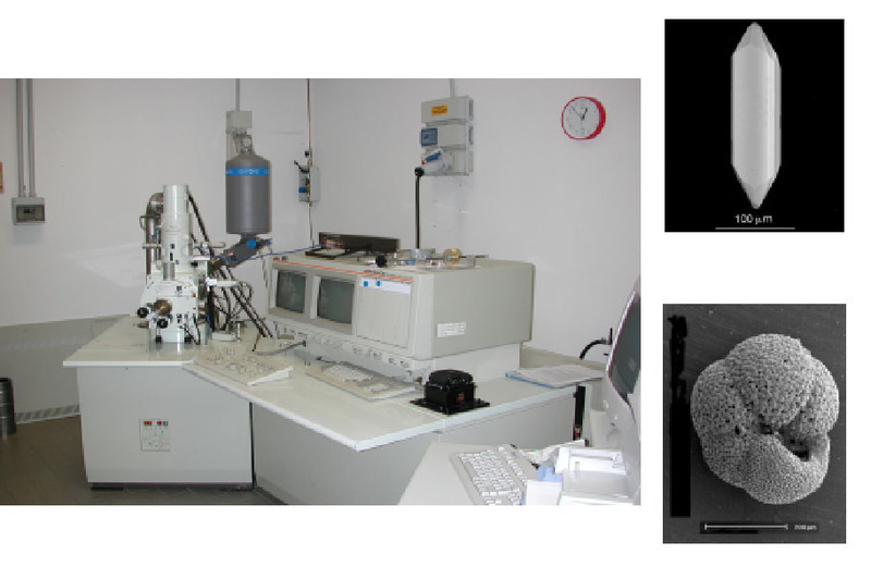
Scanning Electron Microscope (SEM) and Microanalysis Laboratory - research laboratory
Componenti
- Salvioli Mariani Prof.ssa Emma (Responsabile)
- Barchi P.I. Luca (Tecnico)
Contatti
Attività

INSTRUMENTS
- Scanning Electron Microscope JEOL 6400, operating under high vacuum conditions (10-4 Pa) and up to 300.000 magnifications
- Energy Dispersive X-Ray Microanalysis System (EDS) Oxford-INCA, with Si(Li) window-less detector and a friendly software
- A vacuum evaporator JEOL JEE-4X for specimen preparation
POSSIBLE ANALYSES
- Secondary electron images; the acquisition of secondary electron signal allows to obtain morphological information on the sample. The signal of secondary electron is collected by a scintillation detector

- Backscattered electron images; the signal of backscattered electron is collected by a pair semiconductor detector to obtain topographic and compositional information. The compositional image shows the atomic number contrast of the specimen (as shown in the picture below).

- X-Ray detection by Si(Li) window-less detector. It is possible to have the following informations:
- Qualitative analyses on different types of samples
- Standardless semi-quantitative anlyses
- Quantitative analyses after acquisition of international standard samples, consisting of pure elements, simple oxides or simple silicate compositions
- X-Ray maps to investigate element distribution on the investigated sample surface
- Analyses of element distribution along a line profile (linescan)


FIELDS OF INTEREST
Mineralogy, Petrology, Paleontology, Micropalentology, Geology


Cultural Heritage


Applied Petrography, Engineering


Biology, Chemistry


Pharmacology


